
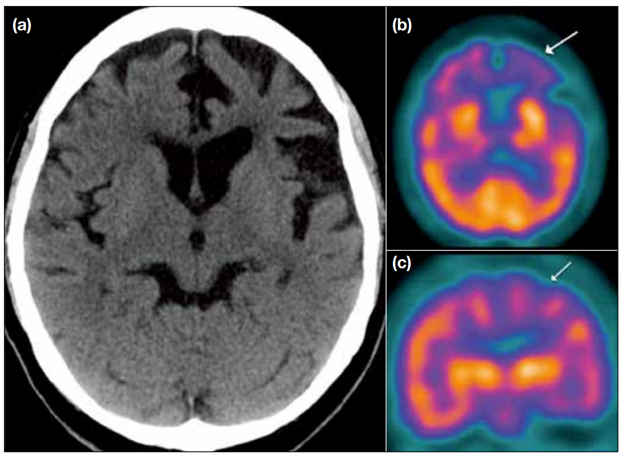
Role of Nuclear Medicine and PET CT in Neurology
August 27, 2021
Dr. Saabry Osmany, MD, FACNM
Written and published by Dr. Saabry Osmany, MD, FACNM
The Deauville scale and Lugano criteria are used for assessment, staging and response of Hodgkin lymphoma (HL) and non-Hodgkin lymphoma (NHL). They both use FDG PET/CT and no other PET tracers.
Deauville score
The Deauville criteria uses a five point scale based on visual analysis of FDG PET/CT imaging of the site of initial (pre-treatment) lymphoma:
| Score | Definition |
| 1 | No uptake |
| 2 | Uptake ≤ mediastinum |
| 3 | Uptake > mediastinum but ≤ liver |
| 4 | Moderately increased uptake compared to the liver |
| 5 | Markedly increased uptake compared to the liver and/or new lesions |
| X | New areas of uptake unlikely to be related to lymphoma |
A baseline (pre-treatment) FDG PET/CT scan is mandatory. Scoring is done pre-treatment and during or after completing treatment.
Lugano Classification
The Lugano classification addresses diagnosis, evaluation, staging, response and follow up of primary nodal lymphomas. Pathology is used to diagnose lymphoma. FDG PET/CT can identify sites for biopsy including discordant disease and suspected malignant transformation. Clinical evaluation criteria are described and included in the classification.
Staging.
FDG PET/CT is used to stage for FDG avid lymphoma and CT for non-FDG avid lymphoma.
Tumor burden
This is calculated at staging and is mainly CT based. This is important as it is used for staging and response, both for FDG PET/CT and CT criteria.
On CT note the six largest target lesions
- These are nodes, nodal masses or extranodal lesions.
- Each lesion should be measured for longest (LDi) and shortest diameter.
- The lesions should be from different regions and representative of overall disease burden.
- The lesions should include any mediastinal or retroperitoneal disease
- LDi should be >1.5 cm for nodes and >1.0 cm for extranodal disease.
The longest diameter and shortest diameter of each lesion is measured in the transverse plane then the values for each lesion are multiplied to give the “product of the diameters” for each lesion. These are then added together to give the “sum of the product of the diameters,” or SPD. This SPD will serve as the baseline for sequential quantification of tumor burden on CT at interim and end-of-therapy FDG PET/CT. The lesions are used for FDG PET/CT follow up.
All other lesions are followed as non-measured disease (eg, cutaneous, GI, bone, spleen, liver, kidneys, pleural or pericardial effusions, ascites).
FDG PET/CT
FDG PET/CT is recommended for staging FDG avid nodal lymphomas particularly before radiotherapy and the classification applies to nodal lymphomas and primary extranodal diffuse large B-cell lymphoma (DLBCL).
The Lugano Classification uses the same scale as the Deauville score but refers to it as a 5 point score (5PS).
The tonsils, Waldeyer’s ring and spleen are considered nodal tissue for staging.
CT
A contrast enhanced CT is recommended for anatomic staging and radiation therapy planning.
Bulky disease
A single nodal mass of 10 cm or greater than a third of the transthoracic diameter at any level of thoracic vertebrae as determined by CT is the definition of bulky disease for HL.
For both HL and NHl record the longest dimension on CT.
Splenic involvement
Best determined by FDG PET/CT uptake even with normal size – homogeneous splenomegaly, diffuse infiltration with miliary lesions, focal nodular lesions or a large solitary mass.
Splenomegaly defined as size >13 cm on CT in the coronal plane.
Liver involvement
FDG PET/CT uptake patterns similar to the splenic patterns.
Bone Marrow Involvement
If FDG PET/CT is performed, a bone marrow aspirate/biopsy (BMB) is no longer required for HL.
In DLBCL a positive FDG PET/CT finding indicates bone or bone marrow involvement but a negative scan requires BMB.
BMB is recommended for all other lymphomas.
Ann Arbor
Based on the criteria discussed a modified Ann Arbor classification is used for staging:
| Stage | Involvement | Extranodal involvement (E) I& II only |
| Limited | ||
| I | One node or group of adjacent nodes | Single extranodal lesion, no nodes |
| II | ≥2 Nodal groups, same side of diaphragm | Limited contiguous extranodal involvement |
| II (bulky) | Meets “bulky” criteria | |
| Advanced | ||
| III | Nodes on both sides of the diaphraphram or
Splenic involvement and nodes above the diaphragm |
|
| IV | Additional non-contiguous extranodal involvement | |
| For HL : A/B – absence (A) or presence (B) of disease-related symptoms | ||
Response
FDG
FDG PET/CT and the Deauville score is used for FDG avid lymphoma and can be used in to assess interim response and/or after completion of treatment and can indicate complete response (CR), partial response (PR), stable disease (SD) or progressive disease (PD). Interim FDG PET-CT is used to assess early treatment response. End of treatment FDG PET-CT is used to establish remission status.
CR, PR and SD FDG criteria apply to the 6 target lesions chosen at initial staging and except for bone marrow (BM) the non-measured lesions are not used in these three possible responses.
CR includes residual masses that are not FDG-avid while increase in a single node indicates progressive disease.
| Response | FDG PET/CT Response Criteria |
| CR | 1 or 2 or 3 (use 1 or 2 for de-escalation cutoff)
With or without a residual mass on CT. No uptake in BM. |
| PR | 4 or 5 decreased from baseline.
No new FDG avid lymphoma. This is responding disease. |
| SD | 4 or 5 stable from baseline.
No new FDG avid lymphoma. No BM change from baseline. Called no metabolic response. |
| PD | 4 or 5 increased from baseline.
New FDG avid lesions, likely lymphoma, with biopsy or interval scan if uncertain. New lesions maybe nodal, extranodal or bony. |
When high FDG physiologic uptake (greater than normal mediastinum and/or liver) is seen in Waldeyer’s ring and extranodal sites with or FDG avid activation is seen spleen or marrow (eg, with chemotherapy or myeloid colony-stimulating factors) this is considered complete metabolic as long as FDG uptake at sites of initial involvement is no greater than surrounding normal tissue.
For PR any persisting focal bone lesions despite nodal response need interval FDG PET/CT follow up, bone biopsy or MRI assessment as bone marrow lesions can take longer to resolve.
A score of 3 from an interim FDG PET/CT scan is considered inadequate for considering de-escalation of treatment.
CT
Used for non-FDG avid or variably avid lymphoma or when PET/CT is not available.
Use sum of product (SPD)
Lesions that split during disease response
When a confluent nodal mass splits into discrete nodes the product of the individual perpendicular diameters (PPDs) of all the nodes should be summed and this represents the PPD of the split lesion. This PPD is added to the sum of the PPDs of the remaining lesions to for response calculations.
If any or all of these discrete nodes subsequently grow, the nadir of each node is used when determining progression (as if each individual node was selected as a target lesion at baseline).
Lesions that become confluent during disease progression
Conversely when a group of target lymph nodes becomes confluent, the PPD of the current confluent mass is compared with the sum of the PPDs of the individual nodes to assess response e.g. a more than 50% increase in the PPD of the confluent mass compared with the sum of individual nodes indicate progressive diseases. The LDi and shortest diameter are no longer needed.
| Response | CT Response Criteria |
| CR | Target nodes/nodal masses must regress to ≤1.5 cm in LDi.
No extralymphatic sites of disease. No non-measured lesion. Organs regress to normal. |
| PR | ≥ 50% decrease in SPD of up to 6 target measurable nodes and extranodal sites
When a lesion is too small to measure on CT, 5mm x 5mm as the default value When no longer visible, 0 x 0 mm For a node > 5 mm x 5 mm, but smaller than normal, use actual measurement The size of the spleen beyond normal (13cm) at baseline decreased by > 50% |
| SD | Target lesions < 50% decrease from baseline in SPD of up to 6 dominant,
measurable nodes and extranodal sites; no criteria for progressive disease met. |
| PD | At least one of the following:
1. An individual node/lesion must be abnormal with: a. LDi > 1.5 cm and b. Increase by ≥ 50% from PPD nadir and c. An increase in LDi or SDi from nadir i. 0.5 cm for lesions ≤ 2 cm ii. 1.0 cm for lesions > 2 cm 2. Existing splenomegaly – the length must increase by > 50% of the extent of its prior increase beyond baseline (eg, a 15-cm spleen must increase to > 16 cm). 3. New or recurrent splenomegaly, must increase by at least 2 cm from baseline. 4. New or clear progression of pre-existing non-measured lesions |
New lesions on CT may be:
- Regrowth of previously resolved lesions.
- A new node >1.5cm in any axis.
- A new extranodal site > 1.0 cm in any axis; if < 1.0 cm in any axis, its presence must be unequivocal and must be attributable to lymphoma
- Assessable disease of any size unequivocally attributable to lymphoma
Hepatic and mediastinal blood pool FDG activity
The Deauville score is based on visual assessment and this is continued in the Lugano classification with no mention of quantitative measurements though these are used in clinical practice. A few approaches described the literature are discussed here.
SUVmax with regions of interest
A group at St. Thomas’ Hospital, London is described as confirming visual evaluation by drawing regions of interest (ROI) around:
- Residual area(s) of uptake, if present,
- The normal mediastinum
- in the arch of the aorta (or just above the aortic root in primary mediastinal B-cell lymphoma)
- avoiding the vessel wall and any areas of calcification.
- The normal liver
- avoiding the edge
- avoiding any individual/single ‘hot’ pixels likely to represent noise
- sampling several axial slices to obtain a representative maximum liver SUV.
These ROIs are used to generate SUVmax which are used to generate the DS.
qPET – target SUVpeak and hepatic SUVmean
This is an approach to semi-quantify the DS by using the SUVpeak of the lesion/residuum and the SUVmean of the liver.
| qPET value | DS |
| 0 | 1 |
| <0.95 | 2 |
| 0.95 to <1.3 | 3 |
| 1.3 to <2.0 | 4 |
| ≥2.0 | 5 |
SUVpeak is the average value in the maximum SUV voxel and the three hottest adjacent ones generated using a semiautomated observer-independent algorithm.
SUVmean of the liver is generated using a cuboid VOI of 30 ml (edge length proportion 2:2:1) positioned in the centre of the right lobe of the liver providing reproducible values regardless of positioning.
The qPET value is the quotient of the mean standard uptake value of the tumour residual SUVpeak and the SUVmean of the liver and the resulting value is translated to the DS as show in the table to the right.
FDG score 5 – Markedly increased uptake compared to the liver
This has been described as > 2 x to 3 x hepatic SUVmax by Barrington et al who also advises timing the post treatment FDG PET/CT scan a minimum of 3 weeks, but preferably 6 to 8 weeks, after completion of the last chemotherapy cycle, 2 weeks after granulocyte-colony stimulating factor (GCSF) treatment and 3 months after radiotherapy.
Summary
The Deauville scale and Lugano criteria are part of an ongoing process to improve incorporation of FDG PET/CT in the treatment of lymphoma. The Lugano criteria incorporates the Deauville scale and provides additional integration with CT, pathology, radiotherapy and clinical practice. Changes introduced by the Lugano criteria include:
- FDG PET/CT is the standard for imaging FDG-avid lymphomas
- CT is indicated for non-avid histologies.
- If FDG PET/CT is performed for HL a BMB is no longer indicated.
- If FDG PET/CT for DLBCL is negative a BMB is only needed if identifying a discordant histology is important for patient management.
- The 5-point scale for FDG PET/CT should be used for response assessment in FDG-avid lymphomas. CT is preferred for non-avid or variable FDG avidity histology.
- Progressive disease by CT criteria only requires an increase in the PPDs of a single node by ≥ 50%.
- Complete metabolic response on FDG PET/CT even with a persistent mass is considered a complete remission.
References
Radiology. 2015 Aug;276(2):323-38. doi: 10.1148/radiol.2015142088.
Imaging for Staging and Response Assessment in Lymphoma.
Johnson SA et al.
J Clin Oncol. 2014 Sep 20;32(27):3059-68.
Recommendations for initial evaluation, staging, and response assessment of Hodgkin and non-Hodgkin lymphoma: the Lugano classification.
Cheson BD et al.
Leuk Lymphoma. 2009 Aug;50(8):1257-60. doi: 10.1080/10428190903040048.
Report on the First International Workshop on Interim-PET-Scan in Lymphoma.
Meignan M et al.
Cancer Res. 1971 Nov;31(11):1860-1.
Report of the Committee on Hodgkin’s Disease Staging Classification.
Carbone PP et al.
Semin Nucl Med. 2018 Jan;48(1):4-16. doi: 10.1053/j.semnuclmed.2017.09.001.
PET/CT for Staging; Past, Present, and Future.
El-Galaly TC et al.
Drug Des Devel Ther. 2017 Jun 13;11:1719-1728. doi: 10.2147/DDDT.S136988.
Lugano 2014 criteria for assessing FDG-PET/CT in lymphoma: an operational approach for clinical trials.
Van Heertum RL et al.
Chin Clin Oncol. 2015 Mar;4(1):5. doi: 10.3978/j.issn.2304-3865.2014.11.03.
Staging and response assessment in lymphomas: the new Lugano classification.
Cheson BD
Eur J Nucl Med Mol Imaging. 2017 Aug;44(Suppl 1):97-110. doi: 10.1007/s00259-017-3690-8.
FDG PET for therapy monitoring in Hodgkin and non-Hodgkin lymphomas.
Barrington SF et al.
Eur J Nucl Med Mol Imaging. 2014 Jul;41(7):1301-8. doi: 10.1007/s00259-014-2715-9. qPET – a quantitative extension of the Deauville scale to assess response in interim FDG-PET scans in lymphoma.
Hasenclever D et al.